Simulation Equipment
Carolinas Simulation Center offers a variety of equipment to help train healthcare professionals to provide care across a continuum of the population. If you have specific questions about our equipment, or you are interested in borrowing equipment to be used outside our center, email us at SimulationCenter@AdvocateHealth.org.
Our equipment includes:
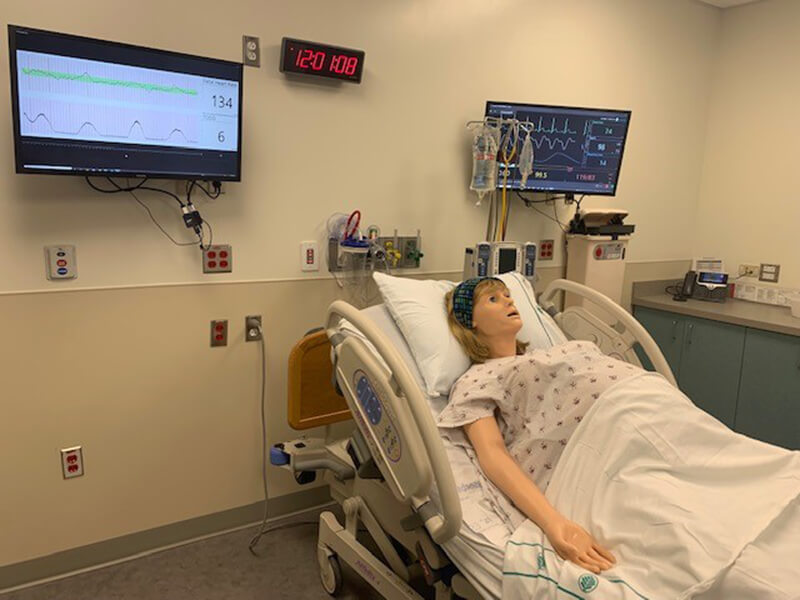
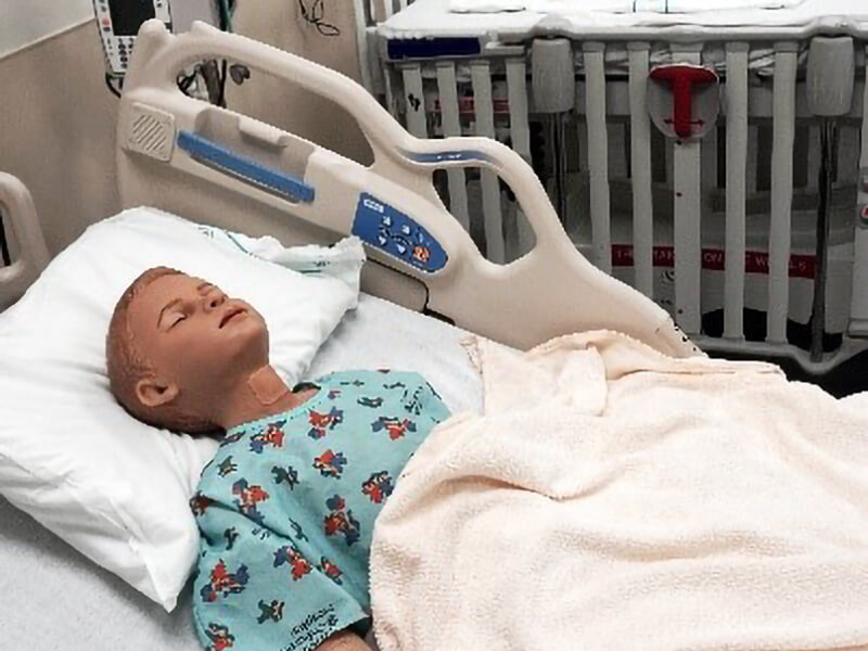
| Patient Simulators | |
|
|
 |
|
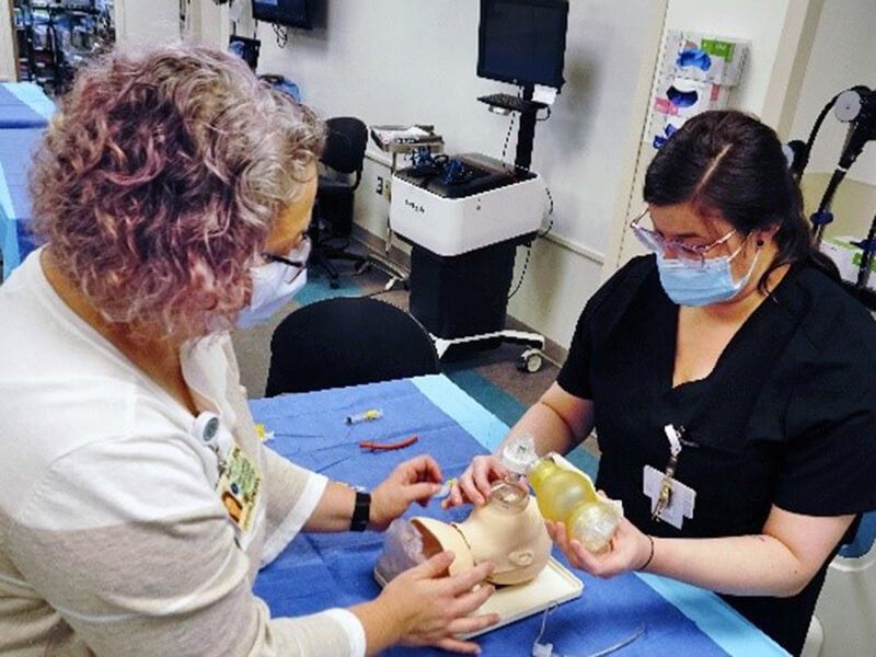
| Task Trainers | |
|
|
|
|
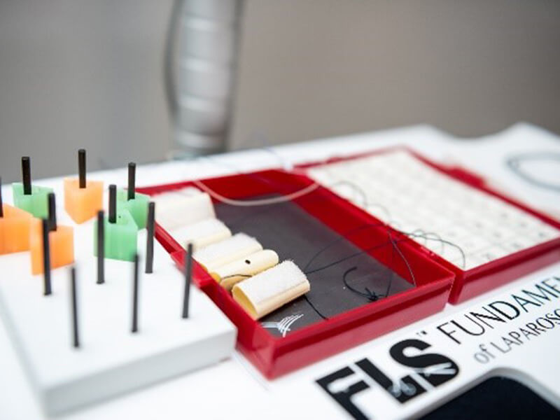
| Virtual and Screen Based Simulators | Clinical Equipment |
|
|
 |
|